

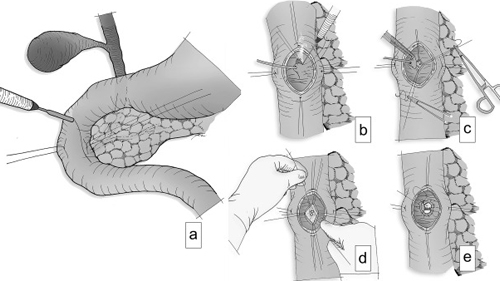
Ampullectomy
Ampullectomy is an advanced endoscopic procedure used to remove selected lesions arising from the ampulla of Vater, where the bile and pancreatic ducts open into the duodenum. In appropriately selected cases, endoscopic removal can provide treatment while avoiding more extensive surgery.
The ampulla of Vater is an important area of the duodenum involved in the drainage of bile and pancreatic secretions. Ampullary lesions may be identified during endoscopic evaluation or investigated because of abnormal imaging, symptoms or suspected precancerous changes.
Dr. Suresh H. Pasiddhi has documented advanced endoscopic expertise, including ampullectomy as part of his therapeutic endoscopy practice.
Conditions
Ampullectomy may be considered for selected ampullary lesions, including:
- Ampullary adenomas
- Precancerous ampullary lesions requiring removal
- Selected early ampullary neoplastic lesions
- Ampullary lesions identified during endoscopic evaluation
- Residual or recurrent ampullary lesions in selected cases
Symptoms
Ampullary lesions may cause symptoms when they interfere with bile or pancreatic duct drainage. Possible symptoms include:
Common Symptoms
- Jaundice or yellowing of the eyes and skin
- Upper abdominal pain or discomfort
- Dark urine or pale stools
- Nausea or vomiting
- Unexplained weight loss or reduced appetite
- Recurrent pancreatitis in selected patients
- Some ampullary lesions may be discovered without specific symptoms
Causes
The development of an ampullary lesion may be associated with several factors, including:
- Development of an ampullary adenoma or other abnormal growth
- Precancerous cellular changes within an ampullary lesion
- Genetic or familial conditions associated with ampullary adenomas in selected patients
- Other abnormalities of the ampulla identified during diagnostic evaluation
The exact nature of an ampullary lesion is determined through appropriate endoscopic assessment, tissue evaluation when indicated and histopathological examination.
Treatment
Endoscopic ampullectomy is performed using a specialized endoscope under appropriate sedation or anesthesia. The lesion is carefully assessed before removal, with additional evaluation or treatment of the bile or pancreatic ducts when clinically required.
Depending on the findings, treatment may include:
- Detailed endoscopic assessment of the ampullary lesion
- Endoscopic removal of suitable ampullary lesions
- Retrieval of removed tissue for histopathological examination
- Assessment of bile duct and pancreatic duct involvement when indicated
- Temporary biliary or pancreatic stent placement in selected cases
- Endoscopic management of residual or recurrent lesions when appropriate
Not every ampullary lesion is suitable for endoscopic removal. Lesions with extensive ductal involvement, invasive features or other high-risk characteristics may require surgical evaluation or another treatment approach.
Prevention & Follow-Up
Regular follow-up after ampullectomy is important to assess healing and check for residual or recurrent tissue.
Helpful measures include:
- Follow the recommended surveillance schedule after ampullectomy.
- Attend planned follow-up endoscopy when advised.
- Review the histopathology report and complete further evaluation when recommended.
- Attend scheduled appointments for removal or exchange of temporary ductal stents when placed.
- Follow post-procedure dietary and medication instructions.
- Seek prompt medical attention for severe abdominal pain, fever, vomiting, bleeding or jaundice after the procedure.
The timing and frequency of surveillance depend on the lesion, histopathology, completeness of removal and individual clinical factors. Long-term follow-up may be recommended because some ampullary lesions can recur.