

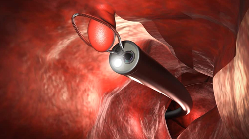
EMR (Endoscopic Mucosal Resection)
EMR (Endoscopic Mucosal Resection) is an advanced endoscopic technique used to remove selected abnormal or precancerous lesions from the lining of the gastrointestinal tract. It allows suitable lesions to be removed through an endoscope without the need for conventional open surgery.
During EMR, the targeted lesion is carefully assessed and lifted or separated from the deeper tissue layers before being removed using specialized endoscopic instruments. The removed tissue can then be sent for histopathological examination to determine the nature and completeness of the resection.
Dr. Suresh H. Pasiddhi has advanced endoscopy expertise, including EMR as part of his areas of interest in therapeutic and third-space endoscopic procedures.
Conditions
EMR may be considered for selected gastrointestinal lesions, including:
- Selected precancerous gastrointestinal lesions
- Suitable superficial gastrointestinal tumors or neoplastic lesions
- Large or flat gastrointestinal polyps
- Selected esophageal lesions
- Selected gastric lesions
- Selected colorectal lesions
- Abnormal mucosal lesions requiring complete endoscopic removal and tissue evaluation
Symptoms
Many lesions treated with EMR may not cause specific symptoms and can be detected during screening or diagnostic endoscopy. When symptoms are present, they may include:
Possible Symptoms
- Blood in the stool or rectal bleeding
- Occult gastrointestinal bleeding
- Unexplained iron-deficiency anemia
- Persistent abdominal discomfort
- Changes in bowel habits
- Difficulty swallowing for selected esophageal lesions
- Unexplained weight loss in some patients
Causes
Gastrointestinal lesions that may be considered for EMR can develop due to different underlying factors, including:
- Abnormal growth of cells within the gastrointestinal lining
- Precancerous changes within gastrointestinal mucosa
- Adenomatous or other polypoid lesions
- Superficial neoplastic changes identified during endoscopy
- Other localized mucosal abnormalities requiring endoscopic removal
Treatment
EMR is performed through an endoscope under appropriate sedation or anesthesia. The lesion is carefully evaluated to determine whether endoscopic removal is suitable before specialized instruments are used to remove the abnormal tissue.
Depending on the lesion and its location, treatment may include:
- Detailed endoscopic assessment of the lesion
- Submucosal lifting of suitable lesions before resection
- Endoscopic mucosal resection using specialized accessories
- Removal of lesions in one piece or in selected cases in multiple pieces
- Retrieval of resected tissue for histopathological examination
- Endoscopic control of bleeding when required
- Follow-up endoscopy to assess the treatment site when advised
The appropriate endoscopic technique depends on the lesion's size, location, appearance, depth and other clinical findings. Some lesions may require a different advanced endoscopic technique or surgical evaluation rather than EMR.
Prevention & Follow-Up
Regular follow-up after EMR is important to confirm adequate treatment and monitor for residual or recurrent lesions.
Helpful measures include:
- Attend scheduled follow-up endoscopy when recommended.
- Review the histopathology report with the treating gastroenterologist.
- Follow dietary and medication instructions after the procedure.
- Complete additional investigations or treatment if advised based on the pathology findings.
- Follow appropriate gastrointestinal screening and surveillance recommendations.
- Seek prompt medical attention for significant bleeding, severe abdominal pain, fever, persistent vomiting or other concerning symptoms after EMR.
The timing of surveillance depends on the lesion, histopathology, completeness of removal and individual risk factors. Regular monitoring can help identify residual or recurrent tissue at an early stage.